Visual Neuroscience Lab
  • Home
  • Neurodesktop
  • Behavioral
  • Labmeeting
  • DICOM to GLM
  • MRI
  • People
  • Internship
  • Other Topics
  • Archive
  1. MRI
  2. Brain Atlas Viewer
  • Home
  • Neurodesktop
  • MRI: from DICOM to GLM Analysis
    • 00 - Overview
    • 01 - DICOM to BIDS
    • 02 - Check IntendedFor field of the fmap’s JSON
    • 03 - T1w for PBn
    • 04 - Defacing
    • 05 - Removing Noise Scan
    • 06 - fMRIprep
    • 07 - Probabilistic Retinotopic Mapping
    • 09 - ROI Analysis
    • Troubleshooting
  • MRI
    • MRI studies: general information
    • 3D printing a brain
    • 7T anatomical analysis on neurodesk
    • MRI BIDS and OSF
    • fMRIprep analysis
    • MNI space
    • Control Masks
    • Old MRI setup
    • Update to the MRI lab setup
    • Physiological noise correction
    • Brain Atlas Viewer
  • Behavioral
    • Vision tests
    • BIDS for behavioral studies
    • Custom-built chin rests
  • People
    • Stipends and prizes for master and PhD students
    • Coffee time
    • Participant compensation
    • Things to do if you are leaving the lab
    • Scientific writing in practice
    • Research funding opportunities
  • Internship
    • Pflichtpraktikum
    • Internship Checklist
    • internship/claustrum_segmentation.qmd
  • Other Topics
    • Calculating a priori sample size using G Power
    • Gamma correction
    • Contribute to the website
    • Older ways to access the server
  • Labmeeting
  • Archive

On this page

  • How to use it
  1. MRI
  2. Brain Atlas Viewer

Brain Atlas Viewer

MRI
Interactive 3D viewer of the left and right hemisphere segmentation
Published

September 23, 2026

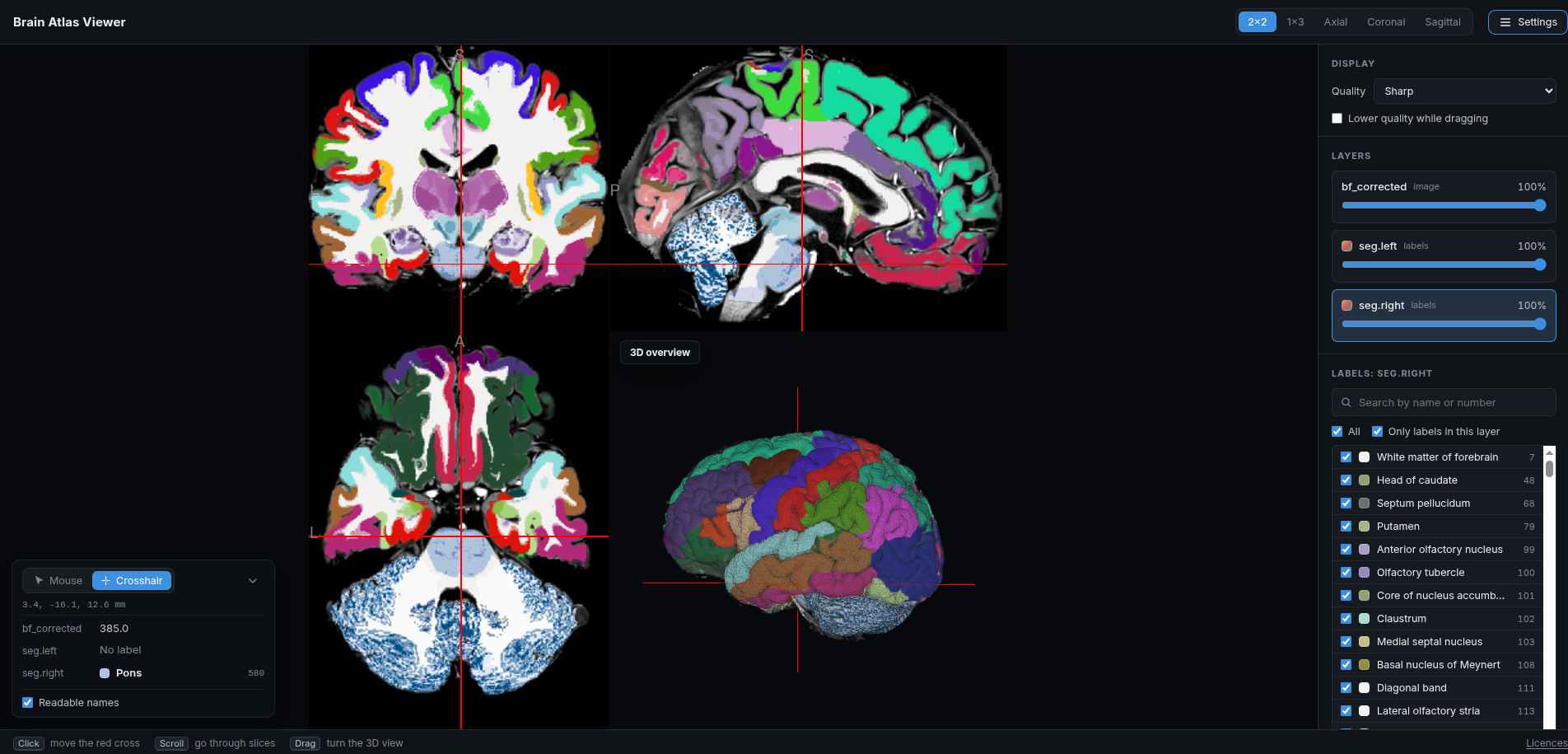
Explore the left and right hemisphere segmentation on the scan, slice by slice and in 3D, and see which structure is at any point.
Open the viewer ↗

The viewer in the 2×2 layout with labels shown

How to use it

  • Show the labels: they start hidden. Open Settings and move the opacity slider of seg.left or seg.right.
  • Navigate: click to move the red cross, scroll to go through slices, and drag to turn the 3D view.
  • Change the layout: the buttons at the top switch between 2×2, 1×3 and a single axial, coronal or sagittal slice.
  • Identify structures: the box in the lower left shows the coordinates and the label under the mouse, or at the red cross in Crosshair mode.
  • Find or hide labels: click a label layer in Settings to list its labels, then search them by name or number and untick labels to hide them.

The viewer opens in a new tab. Use a desktop browser.

To use it offline, right-click the button, choose Save link as…, and open the saved file.

Back to top